Search Results
201 results found with an empty search
- Knee Problems
Most frequent referrals from tumor, infection and non-traumatic problems in the knee region 1. Meniscus tears 2. Cruciate ligament injury 3. Cartilage lesions (chondromalacia, joint mouse) 4. Plika syndrome Knee arthroscopy offers the opportunity to intervene in all of these problems without the need for open surgery.
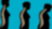
- Multidisciplinary Treatment for Low Back Pain
Multidisciplinary Approach and Treatment in Low Back Pain Physiotheraphy Neurology Physiotherapy Algology Spinal surgery It is possible with the cooperation of physicians working in their branches. Progressive (algorithmic) treatment principles require anti-inflammatory therapy in primary care and bed rest not exceeding 3 days. In resistant and chronic cases, algologists apply block-pain treatments, physical therapists apply physical therapies. All treatments are supported by muscle strength-posture discipline-ergonomic measures with the support of physiotherapists. Surgical compression of neural structures is the last step of choice in recurrent resistant cases of osteoarthritis due to significant loss of disc height. As open surgery indications: Cauda equina syndrome Progressive neurological deficit Failure of conservative treatment Paresthesias that are not obvious but affect life Pain with attacks and requiring rest more than three times a year countable. The indication for minimally invasive (closed endoscopic) surgeries differs at this stage. Prominent neurological deficit, cauda equina may be a contraindication. Endoscopic surgery has a place in cases requiring bed rest more than three times a year, but without an absolute surgical indication. PAIN, which reduces the quality of life and does not respond to conservative treatment, is NOT THE FATE OF THIS POPULATION. It is a treatment that is purely aimed at improving the quality of life.
- Recessive Drug Treatment
We can divide Recessive Drug Treatments into five. 1. Anti-inflammatory Treatment Surgical incisions or skeletal injuries heal with the tissue reaction we call inflammation. Pain is a perception caused by inflammation. We cut the pain and the unwanted effects of inflammation with anti-inflammatory drugs. Steroids are the strongest known anti-inflammatories. We do not use steroids unless it is absolutely necessary. The drug group we use most frequently are non-steroidal anti-inflammatories (NSAIs). 2.Antibiotics We apply prophylactic antibiotic treatment before surgery or for therapeutic purposes due to infection. 3. Antiembolic Therapy During orthopedic treatments, preventive antiembolic treatments, that is, blood thinners, are frequently used because of immobility and the effect of limb circulation. When embolism occurs, treatment is applied with the relevant branch physicians in hospital conditions. 4. Supportive Treatments Elements including protein, collagen and matrix structure needed by the musculoskeletal system are taken orally. Glucosaminoglycans, collagens are recommended orally. Complements the treatments. 5. Conservative Treatments It is the support treatment package that we recommend before the injury to prevent skeletal injury or wear on the cartilages of the person.
- 4th ISMISS Turkey Meeting April 1-3 2011
“Beauty of style and harmony and grace and good rhythm depend on simplicity.” (Plato) We would not be overly ambitious to apply Plato’s maxim to simplifying surgical treatments. To name a few would include successful results of minimally invasive and endoscopic surgeries on failed back syndrome; simple surgical drainage, even on severe diskitis, instead of complicated surgeries; percutaneous endoscopic posterior cervical applications; and contralateral endoscopic approaches. In the ISMISS guide, the term MISS means causing less trauma. The Fourth ISMISS Congress in Turkey continues its objective with the topic, “Minimally Invasive Spine Surgery.” In 2011, the world’s most advanced authorities will once again participate in this event. In this regard, there is now a more imminent preference for endoscopic surgery when desiring less traumatic methods in today’s applications. Making it even more possible to identify disk and foramen endoscopic lesions and treatments are HD camera systems – which heighten human perception multiple times – and greater recording, archiving and classifying potential, with or without various robotic-navigation or targeting systems. Because of spine problems in joint surgery, in addition to macroscopic or microscopic examinations, the necessity of examining without bleeding by using natural gas or liquid must be indicated to observe unattended conditions for identifying the pathologies of tissues. In this respect, new surgical solutions will be accessible through the guidance of our endoscopy ISMISS sessions. Organized on a scientific platform, the sessions will focus on surgical approaches along with multidisciplinary approaches, while encouraging team work and highlighting the philosophy of algorithmic treatment. Each year we have continued to point out innovative studies on minimal invasive surgery. The approved studies will be strongly encouraged during this period. We have also reiterated the principle, Primum nil nocere, or “first, do no harm,” with respect to MISS – which first appeared alongside traditional surgeries but has now moved to the forefront. We invite our colleagues to an academic festive on our scientific platform. Tolgay Satana, M.D. Course Coordinator
- Osteoporosis (Book)
Editor: Prof. Dr. O. Sahap ATIK Authors: Dr. Tolgay SATANA, Dr. Sezgin SARBAN, Dr. Murat A. HERSEKLI Publisher: Meteksan, Ankara, 1998 http://www.tevak.org/?page=books
- What is a fracture? How to Tell a Fracture?
A fracture is a condition in which our bone integrity is impaired. So how do you understand the fracture? You can read about this and other details below. What are the Differences Between Fracture and Crack? Although an unallocated fracture is considered a crack, it is essentially a fracture as both break bone integrity. How to Tell a Fracture? How Is A Fracture Diagnosed? Fracture findings; pain, swelling, excessive movement are in the form of deformity. In cases where there is no deformity, a definitive diagnosis should be avoided without radiological imaging of the bone. Some fractures line a fairly thin line and can be missed, in which case MRI or tomography may be required. Fatigue fractures can only be noticed on MRI. If it is not compatible with conventional x-ray examination, MRI should be considered without hesitation. For fractures that cannot be localized, the location may be determined by bone scintigraphy and diagnosed by localized MRI or tomography. What is the Fracture Treatment Process? Fractures heal with a plaster splint in 6-12 weeks if the contact surface is more than 50 percent, there is no rotation, the angulation is not more than 15 degrees in the direction of movement, the anterior-posterior 10 degrees and is stable during fixation. Non-healing delay exceeding 5 months, nonunion is accepted in cases exceeding 7 months. At Which Stage Is Fracture Surgery Required? If there is no contact between the fracture ends or if it is less than 50 percent, angulation is high and stability cannot be achieved, surgery should be performed. Some fractures (hip neck fracture, muscle attachment ruptures, epicondyle and malleolus fractures) are treated promptly. If muscle and connective tissue have entered between the broken ends, surgery is also performed. What Are The Surgery Options - How Is It Done - How Long Does It Take? In open surgery, the fracture ends are brought face to face and fixation is provided with plates and metals from the bone marrow (intramedullary) long bones from the surface. In rigid and solid fixings, joint movement can be given immediately and weight can be given. If the surgical fixation is insufficient, timing should be done with the surgeon's decision. What is the Recovery Process After the Surgery? Fracture healing is best achieved without surgery. Fracture union is completed in 6-12 weeks with or without surgery. Weight-bearing and joint movement are planned according to the load carried by the bone. Does It Recur After Surgery? If the boiling is not completed, it can be separated from the same place again. The probability of breaking the unbroken bone is the same as the fracture at the same place after union. How Much is the Surgery Fee? It is planned according to the patient's budget.
- Fracture and Dislocation Treatments
A fracture is when a bone is injured by overload or high-energy impacts and loses its integrity. Great forces are required to break the bone except for reasons such as diseases that reduce its strength (Osteoporosis, Osteogenesis imperfecta, cancers and cysts). Fractures caused by the weakening of the bone with diseases are defined as “pathological fractures”. Long and flat bones respond differently to external forces and are injured differently. Long bones are most resistant to loads in the longitudinal direction of transport. Its bending strength is relatively low, but it is weak against shear forces. Although flat bones are more resistant to shear forces, they do not show resistance like long bones during carrying and bending. Fractures; It can be simple (one-piece), segmental and multi-part. If the fracture line comes into contact with the external environment together with skin injury, it is defined as an open fracture. Open fractures are graded according to the size of the wound and the type of contamination. The dirtiest and most difficult-to-treat injuries were reported as agro-manure-related open fractures. In penetrating-explosive injuries such as high-energy or firearms, sudden loads in all three directions cause comminuted fractures. The type of fracture is important in planning the treatment. Simple fractures can be corrected mostly by manual correction (closed reduction) and with plaster-wrap fixation after the fracture ends meet. In cases where the fracture ends cannot be brought together, which prevents the union, the fracture ends are surgically combined and fixed in various ways. If there is a dirty wound in open fractures, surgical cleaning and surgical treatment may come to the fore. Types of Fracture Treatment with Outlines Closed correction and Plaster fixation: Broken ends are manually corrected and brought face to face. It is followed with a maximum angulation of 15 degrees from the direction of movement without at least 50% contact and rotation. While this angulation is considered to be in the upper limits in children, high angulations in adults are directed to surgery since they do not have a chance to reshape. Traction correction: Segmented closed fractures, large bone fractures that cannot be controlled with strong muscles, or spine fractures with dislocation are placed under traction for alignment. Traction is the principle of directly seating the fracture with weights suspended over the wire inserted into the bones. In traction, the ends of the fractures should be brought face to face for up to 72 hours by balancing the muscles. If the procedure is successful, it can be continued until the union is achieved or plaster and other external fixations (external fixator) can be passed. Surgical Methods Closed correction and percutaneous screw-wire-nail fixation: It is the most preferred surgical treatment method today. It can be planned in cases of unstable after fixation, fractures in the muscle-ligament attachment sites, fractures involving the joint, and in cases where movement is required. Open correction and internal-external fixation: If soft tissue enters between the fracture ends and prevents contact, open surgical treatment and fixations are applied in cases of joint-related fractures, growth cartilage fractures, intra-articular fractures and vascular-nerve lesion, in order to remove foreign bodies in open fractures. Metal fixations are not applied in dirty wounds, external methods (fixators) are preferred. Dislocation; It is the deterioration of the relationship of the surfaces forming the joint with each other. Joints are compatible, intertwined sphere-bowl relationship (hip), as well as mostly incompatible structures or structures that do not have sufficient bone coverage. In this way, the joint becomes more mobile but prone to dislocation and vulnerable to external forces. Joint limits are limited by ligament structures, harmony is ensured by meniscus structures and cartilage-like structures surrounding the joint, which we call the labrum. But the most important support is provided by the structure we call the joint capsule. While it provides lubricity with liquid in the joint, it approaches the surface like a suction cup with the negative pressure effect of the closed airtight structure of the capsule and resists extrusion. As a result of dislocation of the joint, forcing the boundaries of the joint, the rupture of the joint capsule, rupture of the ligaments and the structures that provide the joint harmony are destroyed. When the joint is dislocated, it can break the bones that make up the joint. In this case, fracture-dislocation is mentioned. Joint dislocations should be placed in the first 24 hours. It is placed on the joint with special maneuvers closed and fixed with dressing-bandage-plaster. At least 3 weeks of fixation is planned according to the characteristics of the joint. Fractured dislocations, dislocations associated with joint fractures, late-intervented dislocations, dislocations with vascular-nerve lesions and structures that interfere with placement are treated surgically. Whether for fracture or dislocation, the primary goal in modern orthopedic surgery should be prompt detection and early movement. The socio-cultural structure of the patient can also change the way of treatment. Painless, fully mobile joint and healthy limb are the main goals in treatment.
- What is the Difference of Kyphoplasty-Vertebroplasty?
Ky Kyphosis; It is a definition that means hunchback, which includes the conditions that cause the spine to bend forward in the medical language. It is a structural deformity, it may be congenital or as an acquired disease, causing collapse in the vertebrae and may result in kyphosis. Humpback treatment is eliminated by the corrective interventions of spine surgery. Since it is generally an intervention performed at a young age, open surgery covers major surgeries using metal screw and rod systems. Since it is one of the most severe surgeries in orthopedics, surgical treatment of humpback and problems caused by aging was avoided. The method of bone cement injection to fill the gap created by a bone tumor in the spine, first applied by a Radiologist Drummond in 1984, when “osteoporosis = decrease in bone density” emerged as a cause of reducing the quality of life of the elderly population in societies with the increase in the elderly population after the Second World War; The definition of “vertebroplasty” has begun to be used. Osteoporosis patients were disconnected from daily life due to both deformity and pain, and they were lost due to fatal causes, from embolism to respiratory problems due to immobility. Vertebroplasty method not only fills the gap in vertebral tumors that destroy and weaken the bone structure, but also ensures the destruction of the bone tumor with the cement used. The situation is a little different in osteoporosis. Cement is injected using high pressure with vertebraplasty since there is no cavity to be injected although the bone density decreases. Its use has been restricted because the cement injected with high pressure leaks into the spinal canal and reaches the lungs through the blood. During these periods, the idea that balloons used in vascular surgery would straighten the spine of osteoporotic patients in the bone, create a gap and replace it with cement was developed and put into practice. After the first applications were carried out successfully in Chicago, the concept of “Kyphoplasty” became one of the treatment options. The final point reached in the vertebroplasty-kyphoplasty debate, in which physicians performing spine surgery in the last fifteen years question their habits, is that kyphoplasty is safer and its effect on kyphosis correction is significant. However, kifolpasti is ineffective in the treatment of structural humpback. It is effective in vertebral collapse due to fresh osteoporotic fractures and tumors. Although there is still no consensus among surgeons on cement leakage, both methods can be safely applied, respecting the physician’s habit. My personal practice is that kyphoplasty is safer. Finally, we prefer the use of cement as a filling material only in tumor patients. We use organic products such as calcium-phosphate that regenerate bone instead of fillers that cannot be absorbed and destroyed in polymethyl methacrylate content. Although these products are not mechanically very robust, they are not different in terms of efficiency. In addition, it keeps us away from the disasters that polymethyls cause by destroying bones in the long term.
- Trauma and the Skeletal System
Although the concept of trauma is defined as a physical injury that changes the body, it is known to have complex biochemical consequences (see: endocrine response to trauma) in a living organism. The skeletal system provides the first defense in physical interactions with its strong carrier, skull and rib cage-like covering feature. Trauma; Whether blunt, penetrating or post-explosive, the skeletal system acts as a protector. Since injuries that occur in this way are very specific and involve many systems; Concepts and specialties such as trauma center, trauma team, trauma surgeon have developed. Orthopedics and traumatology specialist works as part of the trauma team in the treatment of skeletal system injuries such as fractures, dislocations and crushes that occur after trauma. The aim of orthopedic surgery is to remove foreign bodies with injuries resulting from trauma, to repair fractures and to provide body integrity by placing dislocations. First of all, the vital functions of the patient should be corrected by the trauma team. Joints can be placed during these procedures. In particular, determinations and tractions to immediately remove the pressure and correct the alignment, such as significant alignment disorders that may cause limb loss, such as knee dislocation, and spinal fracture, sometimes have to be performed simultaneously in the ABC rule. For example, a neck fracture may require immediate intervention as it will stop the respiratory center from functioning. The traumatized patient should be intervened by trained personnel, it is a known fact that unconscious interventions, even with first aid, will cause loss of life. When the trauma team reaches the area, after the first procedure, “separation=triage”, critical patients undergo immediate resuscitation (resuscitation). Skeletal system problems are carefully evaluated and necessary interventions such as appropriate temporary fixations and stopping bleeding are carried out immediately. Critical dislocations are seated or placed in a safe position, and spinal fractures are carefully identified and prepared for transport. Whether the transport will be carried out by air or by land, it is planned according to the timing of the intervention and the trauma center is warned. After the patient’s vital functions are preserved and the transfer to the center is completed, the internal organs, thoracic cage and skull injuries are treated immediately and the necessary skeletal structures are determined. The timing of the treatment of skeletal trauma needs to be “immediate” in modern orthopedic surgery. The aim is to prepare the patient for action and to protect the patient against catastrophic consequences such as fat embolism. Fractures that are detected and treated early will heal better and reduce the strain on other organ systems. Otherwise, the body balance will be disturbed by the metabolic load in the trauma body. The best rehabilitation is immediate treatment. Skeletal system surgery was performed without creating new wounds as much as possible, which enabled the development of percutaneous surgeries that kept the patient away from new traumas. Today, long bones can be treated with nails embedded in the bone marrow or with plate supports that stay away from the bone. In the near future, fixation materials that dissolve in the body instead of metal, and even bone adhesives will be put into widespread use.
- Hallux Valgus Surgery - What is Hallux Valgus?
Before details of Hallux Valgus Surgery, let's briefly answer the question of Hallux Valgus - what is the big toe bone protrusion. What is Hallux Valgus? It is the outward curvature of the big toe. However, there are actually three components of the scallop, which include pronation and joint bursitis. It is often accompanied by an inverting of the comb bone (varus). It should be kept in mind that metatarsus primus varus (MPV) causes hallux valgus along with it. Most of the preventable hallux valgus disorders at a young age are MPV. The patient complains of deformity, as well as swelling of the thumb joint and the bad appearance of the protrusion. Since the joint axis is distorted, the joint pains increase with movement and the width is restricted as a result of wear over time. A stiff thumb (hallux rigidus) may develop. Thumb extension disappears. Hallux Valgus Surgery How is Hallux Valgus Treated? Night splints, bursitis treatment, and appropriate shoes may be beneficial before the deformity progresses. Surgical treatment should not be delayed in case of malalignment (metatarsus primus varus, disruption of the joint connection). Medial capsulorrhaphy and lateral release, bursectomy and bunionectomy are applied in soft tissue entrances. Violation of the joint border, which we call the sulcus while removing the bunion, disrupts the joint functions, causing postoperative dissatisfaction and a stiff thumb. In this respect, bunionectomy is very critical and should not be exaggerated in such a way that it does not exceed filing. In metatarsus primus varus where the alignment is disturbed, or in bone curvatures, corrective osteotomies are made on the metatarsal and phalanx bones to correct angular problems and make the joint compatible. At Which Stage Is Hallux Valgus Surgery Required? Surgical treatment should not be delayed in case of malalignment (metatarsus primus varus, disruption of the joint connection). Misalignment should be corrected immediately, joint harmony should be ensured and wear should be prevented. In cases where there is no malalignment, if orthoses to correct soft tissue do not help and frequent bursitis painful joints occur, surgery is a valid solution. What Are The Surgery Options - How Is It Done - How Long Does It Take? It is in the form of soft tissue surgeries and bone surgeries. Soft tissue surgeries; It includes lateral release, medial capsulorrhaphy, and bursectomy. Bone surgeries are osteotomies that correct the alignment. Osteotomies correct bone axis disorders, provide alignment, and create joint harmony. Soft tissue surgeries are also performed along with it. The operation time may vary between 30-90 minutes. What is the Recovery Process After the Surgery? A 3 to 6-week healing period includes the time that special shoes or slippers should be worn. A plaster cast may be required in bone surgery. In cases where bone fixation is rigid, it is possible to press early or to give weight to the heel. However, the possibility of not being able to step should be taken into account. Will It Repeat After Surgery? Recurrence may occur after soft tissue surgeries. New osteotomy is rarely required in bone surgery. How Much is the Surgery Fee? It depends on the type of surgery and the use of materials. Pricing is made according to the hospital class determined according to the patient's budget.
- Spinal Canal Stenosis Closed Endoscopic Treatment
If the spinal canal stenosis occurs congenitally, it requires open surgery at an early age. The canal stenosis that occurs with aging is very different and physiological. After proving that the stenosis of the spinal canal was at the disc level and that there was no canal stenosis at the lamina level, we claimed that there was no need for a screwing (fusion) attempt between the laminectomy used in open surgery and the ring that had to be performed as a result. After proving that the stenosis of the spinal canal was at the disc level and that there was no canal stenosis at the lamina level, we claimed that there was no need for a screwing (fusion) attempt between the laminectomy used in open surgery and the ring that had to be performed as a result. In spine endoscopy, we can easily reach the foramen tract, which we use to remove the lumbar hernia, and open the canal stenosis in a 6 mm hole. In this way, our patients can stand up after 2 hours and get rid of the pain. Moreover, this surgery can be performed awake with local anesthesia without the need for anesthesia.
- Alas I Have a Hernia I Can't Do Sports!
I have a hernia. Can I exercise? According to the data of the World Health Organization, one out of every 4 people has to have bed rest until the age of thirties at least once in their life due to spinal pain (neck, back and waist). Half of those who suffer from spinal pain need medical treatment. Every year, one million of 9 million people who do not respond to medical treatment all over the world need severe surgical treatments (absolute treatment-elimination of the cause), while the remaining eight million seek a solution to spinal pain by restricting their activity with conservative treatments. This situation results in psychological destruction and unhappiness for those who live actively and make daily sports activities a lifestyle, and every year millions of people give up sports, that is, life. I Have a Hernia. Can I Exercise? Why is that? Open surgical procedures are often performed with certain medical indications. No physician will apply an initiative that will not further improve the quality of life of his patient. If open surgery is useless, it recommends activity restriction instead. Benefit-harm ratio is always considered in treatment. The problem is that the old physical activity cannot be guaranteed due to the damage to normal tissues while reaching the diseased area during surgical treatment. Therefore, terminal treatment with classical surgical interventions cannot be applied to the millions of active living people who suffer from paralysis and severe pain that cannot be treated, except perhaps the lucky one million patients. In the last two decades, surgeons trying to reach the relevant segment of the spine without damaging the normal anatomical structures laid the foundations of minimally invasive surgery. Previously, they used a thin steel tube and their tools among dangerous anatomical structures. Initially, the results were promising, but not very bright. However, with the development of endoscopic systems, this time they were able to project the area on the screen and enlarge it, and at the same time treat the affected tissues. Physicians who have been trained by authors such as Hijikata and Kambin over time have made it common to lift their inpatients all over the world by walking from the operating table without pain. Why? Minimally invasive treatment indication is centered on quality of life. Pain that requires bed rest more than three times a year requires treatment because the person may be out of work. Not being able to do sports or restricting daily activities is a reason for treatment, because health by definition; "it is a state of complete mental and physical well-being". There is no health without movement. How? Treatments are applied in sterile surgical settings (operating rooms). Appropriate equipment (endoscopic camera, endoscopic surgical instruments and fluoroscopy - simultaneous x-ray) support, trained spine surgeon should be available. It is imperative that the person to be treated is compatible and not overly obese. Because the intervention is performed under local anesthesia by percutaneous skin puncture through a 0.5-1 cm hole. The patient is fully awake, there is almost no blood loss, he can confirm that he has recovered during the surgery and terminate the procedure himself. It is possible to be discharged the same day or even sunbathe on the beach. Driving for a short time and doing sports can be prevented. However, patients can immediately return to desk jobs. In addition to the low duration of hospitalization and low cost, making the person productive immediately is a great contribution to the country's economy. Where? Minimally invasive spine surgery was born in America and has developed to form academies in Asia and Europe. Among hundreds of physicians who have undergone systematic training today, successful Turkish physicians have also started to implement these initiatives in our country, especially in the last two years. There are only specialized centers in the world for minimally invasive endoscopic spine surgery. In the near future, the centers mentioned with finger in our country will be out of the ordinary. Who? This intervention is applied by spine surgeons who have applied and know the classical methods of spine surgery (Orthopedics and Neurosurgery specialists), who have completed their endoscopic surgery training and have proven that they can perform appropriate minimally invasive surgical techniques with certificates. Do not forget; Medical treatment and recommendations should be made to ensure a better quality, happier and healthier life. Otherwise it's useless. Do you have a hernia, have you been excluded from sports? Do not be afraid there is a cure.