Search Results
201 results found with an empty search
- Joint Health
Painless joint freedom of movement. Stagnant life can lead to unexpected problems such as vascular diseases, internal organ failures, and stroke. The joint moves with the sliding cartilage surfaces. Injury on these surfaces, factors that disrupt the surface also disrupt the opposite side throughout the movement. Over time, both surfaces begin to erode each other. In a small spot, a groove the diameter of the injury appears, the articular cartilage quickly disappears. Joints are ball-slot, hinge structure that works in harmony. The axis of both surfaces must be the same, the covering areas must be the same. We can protect our joint health! Our joints become firmer and stronger as we move. A balanced, protein-rich diet and regular exercise are beneficial. Excessive weight gain should be avoided. A sprained load on the joint may be caused by weakness of muscle strength. Exercises to increase muscle strength should be done. The type of exercise should be appropriate for your level. If you have not walked on a flat road, it is inevitable that you will be injured while running on the treadmill. The fact that the items you use around you are in harmony with you (ergonomic) will reduce your chance of injury. Adapt your surroundings. Accurate diagnosis is possible with examination! When you have a joint injury, you should see an Orthopedics and Traumatology specialist as soon as possible. Radiological examinations performed after the physician’s examination determine the level of injury and the type of treatment. Once the type of injury is determined, the rate of self-repair is understood. Apart from severe injuries, splint-plaster fixations, bandages, therapeutic drugs, painkillers are used that will allow the body to repair itself. Arthroscopic treatment is the gold standard in severe injuries. Don’t let your joint stay still! During injury, recessive treatments may be aimed at restricting the movement of the joint. Immobility of a healthy joint can lead to a decrease in joint lubricity and loss of muscles. Therefore, the treatment is arranged to give action as early as possible. Devices that provide passive movement of the joint surface without weighting can be very effective in treatment. We get help from rehabilitation specialists and therapists for such treatments. Arthroscopic surgery gold standard! Arthroscopy is the gold standard in the diagnosis of delayed healing joint injuries, no matter how advanced and perfect the examination and radiological examinations are. Magnetic resonance imaging may be insufficient for cartilage lesions, plica ruptures and hardening. In this case, intervention is made before joint wear occurs. Today, meniscal tears and ligament ruptures can be treated with arthroscopic methods with excellent results. Correct intervention and stepwise approach are very important when there is a Joint Injury! Your joint may swell after injury, remain motionless and you cannot load. Don’t panic, if you don’t have a real health professional around, a simple bandage, a temporary fix and a cold application is all it takes. Do not be burdened, and rest for a while and contact the health center as soon as possible. Find out exactly what the problem is! You should know what the injury in your joint is, the recovery time and the time to return to normal after recovery. Your doctor will provide information on how joint healing will result. This information can play a key role in explaining your future ailments. Will the treatment affect your daily life and work life? Every person’s daily life expectancy and work life is different. The physician’s decision may vary from recessive treatments to arthroscopic surgical treatments while determining the rest and recovery time. Remember, your joints are the most important part of your life. After the treatment, help may be required to regain the old mobility. In arthroscopic treatments and long angle fixations after severe injuries, we should definitely seek physiotherapy assistance. In simple interventions and diagnostic arthroscopic surgeries, self-exercise may be sufficient most of the time. If it is thought to be inadequate during the controls, it can be included in the rehabilitation program.
- Skeletal System
Without denying Einstein's emphasis, "It is not the skeletal system that keeps man alive, but his beliefs and principles", the skeleton (Skeletal System); We can define it as a system consisting of bone-muscle-joint structures that enables us to stand. Skeleton; Derived from the Greek word skeleton - dried, mummified body. The skeletal system in humans is in the form of "endoskeleton" like most mammals. Some living things, which are invertebrates, contain "exoskeleton - exoskeleton", and when fossilized in this way, they can tell about their appearance in living form centuries later. The skeletal system in vertebrates is like the combination of the skeletons of living things (such as jellyfish, sponges, fish) at different stages of evolution. The human skeleton consists of 206 bones. The bone structure is interconnected by joints that allow movement, supported by ligaments and muscle structures. The main skeleton "Spine" is in the bearing position between the skull (cranium) and pelvis (pelvis) and extends to the coccyx. Spine; In addition to the central nervous system extension in the skull and the carrier feature surrounding the spinal cord, it is also a regulator that ensures the distribution of the nerves. While the surrounding skeleton is connected to the spine by the pelvis below, the more complex ribcage-scapula junction above is connected by a system. The lower peripheral skeleton is related to the muscular and ligament structures in the abdominal region, and the upper peripheral skeleton is related to the rib cage muscles. The main skeletal bones are composed of immobile-semi-mobile joint structures and bones connected to each other. These bones are in flat structures, rich in bone marrow, cell production storage, and contain stem cells. The surrounding skeletal system consists of long bones that are connected to each other by movable joints. The surrounding skeletal joints are complex structures supported by ligaments, meniscal structures, and cartilage. As the joints that provide the movement of the body go to the ends, they get smaller, contain more bones and become more complex; It is specialized as the hand on the upper side and the foot on the lower side. Hand and foot are specialized organs of the main skeleton in the last parts of the skeleton. Today, hand and foot mechanics have not been resolved and their details are little known. Long bones, like the flat bones in the main skeleton, have cell store bone marrow. All bones are a storehouse of calcium, phosphate and some minerals they contain. Bone is a living tissue and acts as an active organ in which the calcium-phosphate level of the body is controlled as well as its carrier feature. In this respect, a healthy body must have a healthy skeletal structure. The cartilage tissue with which the bones are in close relationship consists of cells covering the joint surfaces. It should be remembered that these cells come from the same origin as bone cells before they become specialized in cartilage tissue, and they are structures that can turn into both cells during injury-repair. The boiling tissue that occurs when the bone is broken has a similar character to cartilage and can turn into bone (abundant oxygen, pushing force) or cartilage (less oxygen-pulling force) tissue under different conditions. As a result, the skeletal system is a large tissue, even an organ, actively participating in body metabolism, as well as the connective tissue feature, which is a carrier.
- Endoscopic Lumbar Spine Surgery - Nucleoplasty / Basic Training Course
Endoscopic Lumbar Spine Surgery - Nucleoplasty / Basic Training Course Date: April 26-28, 2019 Venue: Istanbul International Spine Endoscopy Academy Dr Farhad B. BAMARNI Dr Hayati AYGÜN Dr Hikmet ULUĞ Dr Tolgay SATANA Dr Tunç KOÇ
- Minimally Invasive Spine Surgery Applications (Book)
Foreword by Translation Editors If the patient satisfaction of surgical treatment, which remains indispensable among modern medical treatment options, is less than expected, it may not be realistic to question the reasons. In this case, even if an equal distribution is made in the Patient-Surgeon-Treatment triangle, the morbidity of the treatment is always found to be higher than the recessive methods. What should be the ideal treatment in spine surgery? Instead of waiting for the answer to the question to be solved within the same triangle, it would not be misleading to take Hippocrates’ motto “do no harm first” as a guide. Reducing the morbidity of the surgery can be possible by preserving the anatomical integrity as much as possible while reaching the goal and performing the repair duly during the closure. The habit of reaching the target with laminotomy and flavumectomy in disc surgery has been ongoing since 1913. This means that irreversible anatomical deformation during surgery cannot be corrected during closure. Although it is possible to reduce the radicular pain symptoms of the patient, chronic low back pain may inevitably create new problems. Although Pool of 1937 tried to perform surgery with an illuminated tube, it did not contribute to reducing the morbidity of the method. In 1920, Eugen Bircher published the first diagnostic applications of arthroscopy in joint surgery. Although Bricher attempted a thoracoparoscopic treatment, this form of treatment was neglected until the early 1970s. The habit continued with Mallis in disc surgery. In 1955, Mallis used a microscope to reduce damage to anatomical structures and greatly reduce postoperative fibrosis. However, changing the path or method to achieve the goal is the most rational solution. Concurrent with the resumption of arthroscopy in early 1980, Hijikata, Asher and Kambin described the transforaminal safe triangle in the intradiscal treatment pathway for disc surgery. Kambin safe triangle has been published and the concept of “spinal arthroscopy” has been defined. When the first applications were made, and among the confusion of concept and definition, the 1990s was just beginning when special instruments were designed for endoscopic disc surgery. Yeung, Leu and Knight systems had taken their place in the markets by the Wolf, Storz and Endospine companies, respectively. Concurrently, the American minimally invasive spine academy started regular magazine publications, which laid the foundation for the book we are holding. The book was first published in 2000. It was a review of leading publications published in the Minimally Invasive Spine Surgery Journal. However, its most important feature was that it had a multidisciplinary structure. It resembled a consensus text that included all the elements on which minimally invasive surgery was based. Endoscopic surgery was spreading rapidly. While Kambin students were creating the academy, they started reporting successful treatments in Europe and Korea. When the preparation of the new edition of the book became inevitable, a resource book in the form of a textbook was obtained in 2005, without the pioneers taking the background. Martin Savitz was no longer there, but John Chiu was holding the Miss flag, Benjamin Alli was keeping the academy afloat. In our country, there were no specialized sources other than the special issue we published in the Imlas Congress and Joint Disease Surgery magazine, which was held for the first time in 2005. When we held the Ismiss congress in 2008, the congress book was published in English like before. We raised this issue with American Academy members and John Chiu, whom we received support before the second Ismiss congress. brought the source and got the permission of the Turkish translation of the source. Although the work that started in December was a short time, it was concluded quickly and the book was brought to our language. Our country has never lagged behind the world in medical treatments. While minimally invasive surgery is considered as the surgery of the future, when it is indispensable tomorrow, we have no doubt that the best practices will be applied in our country. In this context, we consider the creation of this book a sacred duty. Therefore, we owe a debt of gratitude to my colleagues who accepted the translation and did not spare their assistance, to the employees of Güneş bookstore in the person of Nuran Karacan, and to our congress operations manager Yeşim Tanrıverdi. Dr. Tolgay Şatana To Buy The Book: Nobelkitapevi.com Akademisyen.com Tercihkitabevi.com
- Priformis Syndrome
What is Priformis Syndrome? The priformis muscle is the weakest of the external rotators of the hip. However, since it becomes active especially during hip flexion, it can be easily injured by encountering greater loads than other muscles. Priformis injury can create the typical sciatic nerve area (drop foot and loss of strength in toe elevation) in the acute phase. This situation is temporary. When the priformis edema decreases and the pressure disappears, it can disappear after 2-3 weeks of rest. In chronic cases, the priformis hardened and thickened. By pressing the sciatic nerve just below it, this time it causes pain that does not decrease at rest. What are the Symptoms of Priformis Syndrome? Pain that increases when standing for a long time Weakness in the leg Difficulty walking Pain and numbness radiating to the back of the leg Pain in the hip and coccyx How Is Priformis Syndrome Diagnosed? Diagnosis is difficult, pressure may not be revealed by MRI. EMG can also be confused with root compression, even if there is a herniated disc surgery. Most patients mention sciatica pain that does not decrease despite previous lumbar hernia surgery. In this case, the priformis should be loosened and the sciatica trapped in the canal should be relieved. The patient's complaints decrease and the nerve begins to repair itself. Although surgical treatment is mostly an open surgery, we perform the procedure with endoscopy through a 1 cm hole. Percutaneous endoscopic priformis release is applied under local anesthesia under operating room conditions.
- Waist Health - Athlete Waist Health
We talked about important information for athletes in our pleasant interview with dear Ebru Eryener on waist health. How should we protect our waist while doing sports? First of all, let's assume that the building disk is the most injured when we say waist. Lumbar disc bursts after 400 newtons is loaded and the disease we call hernia occurs. It is one of the most serious injuries. The occurrence of this injury is dependent on the burden. The vector of the load-load changes depending on variables such as the force arm. Briefly, bending, axial loading and rotation are the conditions where the most load is placed on the disc. Strong muscles, correct alignment and correct movement reduce the load on the disc and the risk of injury is virtually eliminated. Therefore, it is of great importance to load a weight proportional to muscle strength, to do the movement correctly, to avoid rotation at the same time during loading. It should be known that the risk of injury is higher in combined movements (turning, bending and loading). During sports, I have students who say that my back hurts when doing movements such as kettlebell swing, superman, cat camel. (Although they do the movement correctly) and there are those who are afraid of this pain and do not want to do the movement. What pain is a sign that the back muscles are working in such a situation? And in what kind of pain should we really be afraid? First of all, the person should not have an alignment problem How do we know that? Hunchbacks, one shoulder low, meaning people with poor posture are at risk. In such athletes, standing movements such as kettlebell, swing or superman should not be given at the beginning, first the core muscles on the ground should be strengthened and in the second stage, the posture should be corrected a little and complex exercises should be started. Although there is no alignment problem in elite athletes, the duration and type of pain is important. If it does not decrease with rest or does not improve with weight reduction, this should not be done more than a muscle regeneration pain. Recently, I have encountered herniated disc in many people. Most of them are very advanced in the initial stage, although some do not require surgical intervention. What are the exercises that you would recommend and never do to these two groups? I would be glad if you explain this in a language that most of us can understand, not as described in medical language. We recommend that patients with lumbar hernia do sports. The back and abdominal muscles should be strengthened and the body-thigh stability should be ensured. We should avoid standing exercises and be content with floor exercises. It should be squat-assisted or it can be on the wall with the ball. If the patient who has undergone surgery has had a closed lumbar hernia surgery as I did, he can go to sports in 3 weeks. You can start on the floor and slowly do the same exercises with healthy people in 3 months. In open surgery, it takes 6 months to return to the gym (sports). Today, people who work at a desk are experiencing back pain and this has become routine. What can you recommend for people who work intensely and experience these pains? Desk workers are a complete catastrophe. There are all of the above. All of them have alignment problem, their muscles are weak, floor exercises must be at the forefront. Since their aerobic capacity is low, such people can be given heart rate tracking and then combined movements. A difficult group that lasted for months, good luck. There is also a stiffness of the waist that happened to me and that I encountered in a few other people. Why does this situation, which is very difficult and restricts the person to move, occur? Will it cause a hernia in the future? And is there anything we can do to avoid this? Your situation may be the type of injury we encounter in elite athletes. Generally, stresses are not on the disc but on the muscle groups carrying the spine (such as kettlebell swing, rupture), muscle-bone adhesion site enthesopathies. (Enthesopathy: It is an inflammatory or degenerative condition that occurs in the areas where the tendons attach to the bone.) The most difficult to heal enthesopathies. While in the other group, they can return to sports with exercise restriction and supports, while enthesopathy may last for months. The simple thing to do is to reduce the repetitions and weight of the injuring exercise and try to continue the exercise by preserving muscle mass and joint width, if not, the resting interval should be increased. My last question is a situation with my mother. One of the vertebrae in the lumbar region is congenitally missing. Would such a situation create a problem in the long term and training? In the case of sacralization, the load on the disc will increase because the force arm is shortened. In this type of athletes, the back and abdominal muscles should be extremely strong, in addition to being supported by the muscles we call spino pelvic (squat basic movement), soft squats can be done after floor movements. It is even possible to do deadlifts within months. An athlete who does a good deadlift will never get injured. The important thing is that regardless of the weight, it is close to the body axis and the shorter the force arm, the closer the center of gravity to the barbell, the less load on the disc. Balanced grip, opening of the feet is important. If one of them fails, the risk of injury will increase. It is said that the shuttle and crunch movement causes long-term lumbar hernia. Because the spine has a bending life. And every time we do this bending movement, it is said that we get a little closer to the hernia. I want to know your thoughts on this subject? Partially correct. With weak muscles, the load on the disc increases and even injury may occur. Disc wall repairs itself in mild injuries. We have a self-regenerating and healing mechanism. However, if the loading is done without full recovery, there may be a hernia from the weakened place.
- Ankle Arthroscopy
Ankle arthroscopy is of great importance especially in chronic foot and ankle pain. Ankle arthroscopy is of great importance especially in chronic foot and ankle pain. What is Ankle Arthroscopy? It is to reveal the problems by imaging the ankle joint with an endoscopic camera (arthroscope) (diagnostic arthroscopy) and the application of simultaneous treatment. Ankle anterior and posterior entrances often include impingement syndromes (ankle impingement), treatment of cartilage lesions, free fragments (joint rat excision), Achilles tendinitis, FPL tendinitis, Haglund's disease, ankle joint restriction treatments, joint membrane inflammation (synovitis-synoyal tissue). hypertrophy and tumors), tumors can be biopsied. Arthroscopy imaging is the gold standard for revealing chronic joint pain that cannot be detected on MRI. It provides a definitive solution for joint pain and stiffness that do not respond to drug therapy. In Which Situations Is Ankle Arthroscopy Preferred? Anterolateral-Anteromedial-Anterior impingement, Posterior-posterolateral-posteromedial impingement: It is the condition in which the contraction of the bone (osteophyte) and soft tissues (ligament thickening-fibrotic healing tissues, synovial thickening) in the anterior part of the ankle erode the cartilage structures. Articular rat is the presence of a free piece of cartilage within the joint. It may not be viewed radiologically. Free fragments within the joint can be removed arthroscopically. In case of delay, it erodes the joint surface. Achilles tendinitis-Bursitis-Haglund's disease, determination of the causes of chronic tendinitis at the back of the ankle, and bursa thickening or bone growths can be treated. Flexor pollicis tendinitis is one of the chronic hindfoot problems. The problem is solved with arthroscopy. With foot - ankle arthroscopy, enthesopathies such as Sinus tarsi syndrome, Plantar fascia problems (epin calcanei) can be detected and treated. If not intervened early, permanent joint damage will occur as a result of cartilage wear. Joint cartilage is lost, joint movement limitation is added to chronic pain and function is lost. In this case, the joint is frozen (arthrodesis) or a prosthesis is applied. How Long Does Ankle Arthroscopy Take? Arthroscopic treatments take 30-45 minutes, reconstruction surgeries that require repair may require more than an hour. After Ankle Arthroscopy? After arthroscopic treatment, the joint is bandaged and cold applied. If the joint is unsupported or repaired on the same day, it can be pressed with partial weight. Movement of the joint is started immediately, but depending on the type of surgery treatment, activity limitation for a few days and rest periods of up to 3 weeks for sports activities may be applied. Usually the weight is given immediately, it is recommended to return to work and sports early with full movement. What is the Recovery Process After the Surgery? Since open surgery is not applied, the wound healing process is very short. After 5 days, the wound holes (5 mm) are completely closed. It can be pressed immediately after surgery. Exercises begin immediately. Joint range of motion is gained immediately. It is recommended to wear shock absorbing sports shoes. Since the soft tissue recovery is completed in 3 weeks, heavy sports activities are not allowed before a month. Will It Repeat After Surgery? In arthroscopic treatments, the tissues that are removed or cut are not replaced. It is possible for fibrotic healing tissues to regrow. These can be prevented with early movement and exercise treatments. In arthroxopic repair surgeries, recurrence is not expected if there is no new trauma in such a way that the repair area is damaged without adequate recovery. Intra-articular injections and PRP combination may be required in joint cartilage abrasions. How Much is the Surgery Fee? It depends on the type of surgery and the use of materials. Pricing is made according to the hospital class determined according to the patient's budget.
- Shoulder Impingement Treatment
Impingement Syndrome Treatment Before giving information about Shoulder Impingement Treatment, let’s answer the question “What is Shoulder Impingement”. Impingement-impingement syndrome is a condition that results from the narrowing of the acromioclavicular bone-ligament complex, which surrounds the joint where the shoulder rotator muscle group and the burs are located, and the space between the muscle (subacromial space). Previous innocent shoulder traumas, recurrent shoulder dislocations, subacromial space in people working on their head; The bursa thickening is narrowed by osteophyte protrusions in the bones, it is known that the inflammation (inflammation) that develops with the injuries of the muscle structures in this space improves compression with the healing tissue over the years. How to Treat Shoulder Compression? In shoulder compression, the bony protrusions under the acromion that narrow the subacromial space are cleaned and leveled (acromioplasty), the simultaneous thickening muscle-bone cushion is removed (bursectomy), if any, muscle tears are repaired (rotator cuff repair). Drugs such as cortisone should not be administered after or before these procedures. In Which Situations Is Arthroscopy Preferred for Shoulder Impingement Syndrome? Arroscopic decompression is the gold standard in shoulder compression. Rarely, an open surgery option may be considered. Arthroscopic treatments are at the forefront in modern orthopedic surgery. How Is Shoulder Impingement Arthroscopy Performed? In addition to standard shoulder arthroscopy entry holes, a new subacromial portal is used. After bursectomy, the bone is leveled and muscle tears are checked. It is standard to control joint structures by entering the joint together. How Long Does Arthroscopy Surgery Take? It takes 30-60 minutes. What is the Recovery Process After the Surgery? Action begins immediately. If there is a rotator muscle tear, a rotator muscle tear protocol is applied. Will It Repeat After Surgery? It is not possible, as its recurrence may occur with inflammation that has occurred over decades. How Much is the Surgery Fee? It is arranged according to the patient’s budget. You can contact our doctor for more information about your shoulder pain and the treatment of shoulder impingement syndrome. You can subscribe to our YouTube channel to follow our videos on the subject.
- Our Freedom on the Court is Possible with the Conscious Sports Habit
Perhaps the best part of tennis is that it can be practiced at any age. While trying to get away from the pressure of busy work life, we must question whether our musculoskeletal system is ready for the physical strains encountered on the court. Otherwise, problems start in our musculoskeletal system by trying to do sports. Firstly; Is our musculoskeletal system ready for new loads? Is our endurance enough to return to sports or participate in tournaments? Do we practice and attach importance to enough before we get started? We have to ask ourselves many questions like. We should know well that Endurance is an attribute that we will gain with practice over time. Exercises to increase the strength contribution of fibers with different properties in the muscle mass require regular training. As the athlete regularly participates in the exercises, the endurance will develop automatically. You have been doing sports for a long time, but if you have exceeded your stay on the court a little that day, you have an unbearable heel pain, you have become almost unable to step, you should start the treatment by paying attention to your shoes. If the court floor is not soil or grass, the usual tennis shoes on the hard ground will cause the abnormal loads of the under-heel fat pad to reflect directly on your heel bone. This situation may cause heel pad compresibility synd. You should choose shoes that have shock absorbing structures at the sole and that grip the heel. Our foot transfers our weight to the floor at three points. These are the protrusion under the heel bone (calcaneus), the end of the comb bone of the 1st and 5th fingers. The hard floor will cause pain in the normal foot by exerting pressure effect on these points. The second problem may be dynamically disturbances of the common beams of the inner foot muscles (plantar fascia). The common beam plantar fascia provides movement with the spinning wheel mechanism. Plantar fasciitis is when muscle groups with insufficient endurance give the load before the adhesion and cause problems such as tearing. Although this situation requires a physician’s intervention, it once again reveals the importance of shoe selection. Let’s not forget that the famous Achilles tendon is attached to our heel bone. It attaches to the heel as the common beam of the muscles that start from our Achilles leg, performing the function of jumping and walking. In cases where the durability is low, loading may cause similar breaks. If the loading occurs to the place of direct adhesion, it is “insertional-adhesion” tendinitis, above it noninsertional. In these two cases, whose treatments are different, your physician’s guidance will be required. Finally, if we touch on the problems our joints will encounter on the court, torsions can injure ligament and cartilage structures. Since strong muscle structures will provide balanced weight transfer, the risk of joint injury is minimal. When the joint limit is forced, respectively, our capsule, our ligaments, then our bones resist the forces. In a balanced load, ligament rupture, fractures and more importantly, cartilage injuries do not occur. Let’s not forget, our freedom on the court depends on our technique, our endurance, as well as our musculoskeletal health. Do not hesitate to consult a physician for foot pain that does not go away despite changing your shoe preferences. Healthy sports. Note: This Article is the text of the article I wrote for the Sports International magazine.
- Endoscopic Disc Surgery
It is possible to get rid of hernias and canal stiffness on the same day !!! It is the process of removing the neck, back and lumbar hernia without anesthesia and without stitches using a 5-7 mm camera. ENDOSCOPIC SPINE SURGERY IS A PROPER AND SENSITIVE PROCEDURE This operation, which provides great comfort to the patient, can also be performed under local anesthesia. The patient is laid face down. By opening a 6mm hole in the skin, the disc and herniated part is reached directly through the camera. SELECTIVE ENDOSCOPY Endoscopic surgery preserves the tissues, the part that presses the nerve is removed without cutting any muscle and touching the bone structure and ligaments. CHANNEL THICKNESS AND CLOSED ENDOSCOPY The endoscopic camera displays all the structures that compress the root nerve as a result of canal stenosis and allows the nerve to relax and get rid of pressure, and at the same time, MRI can even solve problems that cannot be seen. DISCOGRAPHY SURGERY is done by piercing the skin. Disc with a needle inserted through the foramen hole the inside is painted and the hernia is detected. This method is called Discography. When there is more than one hernia, the level to be intervened can be determined by discography. SELECTIVE ENDOSCOPIC DYSCECTOMY (DISCOTOMY) The hernia is directly reached through the tube placed over the needle. With the instruments passing through the tube in the camera, only the herniated part is removed. This process is therefore discectomy, not removal of the disc. CHANNEL THROTTLE The real cause of pain caused by canal stenosis due to aging is the foramina where we insert the endoscope. At the same time, the problem is solved by enlarging this hole where the root nerve comes out. There is no need to insert any screws or cages.
- Hip Arthroscopy Surgery - What is Hip Arthroscopy?
Before details of Hip Arthroscopy Surgery, let's briefly answer the question of what is Hip Arthroscopy. What is Hip Arthroscopy? It is to reveal the problems by imaging the hip joint with an endoscopic camera (arthroscope) (diagnostic arthroscopy) and the application of simultaneous treatment. Impingement syndromes due to Femoro-acetabular incompatibility (Femoro-acetabular impingement = impingement; FAS-FAI), treatment of cartilage lesions, free parts (joint rat excision), bursitis, Snapping, repair of fascia tears, labrum tears repair, Perthes cheliectomy, Biopsy can be performed for cleaning osteophytes, joint membrane inflammation (synovitis-synoidal tissue hypertrophy and tumors), tumors. Arthroscopy imaging is the gold standard in revealing chronic joint pain that cannot be detected on MRI. Provides a definitive solution for joint pain and stuttering that do not respond to medication. In Which Situations Is Hip Arthroscopy Preferred? Femoro-Acetabular Impingement = Impingement (FAS = FAI): Painful joint movement limitation and abrasion resulting from femoral head disharmony of the hip joint's acetabulum (joint cavity). There can be three types of incompatibility: 1 - There can be three types of incompatibility: 2 -If the structure of the head is larger or different in size than the joint cavity, or its axis is different and its shape is elliptical, the wedge = CAM lesion develops and tears the joint edges 3 - Combination of these two situations (combined = mixed type) Labrum tear:The acetabulum is a structure that stabilizes the joint in the labrum cartilage structure at the edge of the joint cavity. In case of tearing, pain and stiffness occur in movements that force the joint limits. It is repaired closed with hip arthroscopy. Snapping is the displacement of muscle beams in the form of painful jumping during contraction and movement as a result of coming out of their grooves or losing their support. With hip arthroscopy, the cause is repaired, muscles and tendons are fixed.r. Bursitis: It is the swelling and inflammation of the cushions between the muscle gates. Bursectomy is applied with arthroscopy. Synovitis:It is inflammation of the joint membrane or tumoral inflammation. The synovia can be taken by arthroscopy and pathological examination can be performed by biopsy. How Long Does Hip Arthroscopy Take? Since hip arthroscopy requires special operating room conditions, it is one of the operations whose preparation takes a long time. Arthroscopic procedures can take up to 1-2 hours. What Should the Patient Do Before Hip Arthroscopy? Load can be given immediately, except for postoperative repair operations. In some of the repair operations, it is possible to give weight early. Depending on the strength of the suture material used, it can be pressed immediately or after 3 weeks. After the surgery, joint movements are started immediately and the movement limits are tried to be reached. Exercises should be extremely regular and diligent. What is the Recovery Process After the Surgery? Wound healing is completed within 5 days, if stitches are made, 5 mm wounds are immediately closed and a bath can be taken. Depending on the type of surgery, it is immediately mobilized and the patient returns to work. The rest period may vary between 3-6 weeks for athletes or professionals working in jobs based on physical force. Will It Repeat After Surgery? In arthroscopic treatments, the tissues that are removed or cut are not replaced. It is possible for fibrotic healing tissues to regrowth, these can be prevented with early movement and exercise treatments. In arthroxopic repair surgeries, recurrence is not expected if there is no new trauma in such a way that the repair area is damaged without adequate recovery. Intra-articular injections and PRP combination may be required in joint cartilage abrasions. How Much is the Surgery Fee? It depends on the type of surgery and the use of materials. Pricing is made according to the hospital class determined according to the patient's budget.
- Minimally Invasive Surgery as the Concept of God Physician Ends
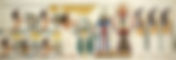
Even twenty years ago, medicine and physicians, which were expected in front of them with endless respect and whose post-treatment debt of loyalty could not be paid with expensive gifts and money, do not exist anymore… In fact, it would not be wrong to say that everything started with Hippocrates… The father of Egyptian medicine, Hermes Trismegistus, the representative of Toth’s teachings on earth, drew the surgical borders of Imhotep in 460 BC in Kos in Anatolia. While the principle of “first do no harm – primum non nocere” instilled the cult of Hippocrates in the Anatolian people, the option of treatment with drugs and the demonstration that abscesses can be healed without causing wounds brought surgical treatment to a level that almost made them forget. Then, in the centers of Pergamon built with temple architecture, medicine ceased to be a kind of alchemy-sorcery, and treatment of diseases was planned with certain formulas, as today’s medicine accepted as reference. In these years, medicine still retained the title of divine wisdom, because recognizing the disease and choosing the right Galenic preparation required an intuitive ability as well as knowledge. The accuracy of diagnosis by using this intuitive ability of medicine is still valid today, but the last fortress in divinity was broken by computer technology, “wisdom for effective accurate diagnosis”. The newly developed computer-based generation can easily reach from disease symptoms to diagnosis and from there to the most accurate treatment option… Yes, physicians are not gods, they are even technicians, moreover, they are too flawed to need their knowledge… This profession group is no longer able to diagnose without engineering miracles. becomes defective and helpless. Advanced electronics, navigation, robotic technologies are about to add nanotechnological devices, in which case it seems not too far for the physician to be replaced by mechanical robots that take more information, go to diagnosis using the genetic code and do this with a small sample of palate-salivary fluid without the need for examination. . Our genetic code is soon to be deciphered. Considering that a genetic defect is responsible for almost all diseases, it is not necessary to be psychic to predict that treatments will descend to the cellular level. Many developments follow one after the other, from cellular level treatment options, from the widespread use of “stem cell-stem cell” therapy to organs obtained by cloning, to nanotechnological pharmaceuticals that are effective at the cellular level. Although it dates back long before stem cell cloning, it has had its share of ethical debates. Discussions about the ineffectiveness of the treatment have almost reached the level of forgetting the bone marrow treatment, which is used extensively in blood cancer treatment. I think that it is a waste of time to look for the ethical basis of transforming the cell into another cell, or even into tissues formed in advanced cells such as nerves, cartilage, and muscles that do not have the capacity to regenerate. While the ethical foundations of genetic treatments, cloning and stem cell treatments were rapidly established, the increase in high revenues from ‘off-shore’ treatments was inevitable and impossible to control. Classical medicine has been in the process of extinction as long as it has received different trainings apart from a classical medical education and did not include trainings that have mastered engineering technologies. The name Asklepion, which was divided by breaking his divine staff long ago, has become unforgettable. It seems extremely risky to leave technologies with a high learning curve, such as endoscopic devices, which require a lot of skill to use, with or without the development of navigation technologies in recent years, only to physicians who have received classical medical education. While the thought that it would be more accurate to practice medicine with an ultra-modern robot, all under the control of a good medical engineer, becomes widespread, I involuntarily take a nostalgic glance at our diplomas on the wall… While divine medicine ended thousands of years ago, in these years when classical medicine came to an end, minimally invasive surgery with less trauma was defined in surgery, where Hippocrates’ expectations with endoscopic technologies have fallen short for now. With this method, which we have been using for decades, not only is the surgical wound smaller, but surgical trauma is reduced and magnified with optical devices, as well as the ability to perceive far beyond the limits of the human eye is gained. Even in this moribund period of medicine, “minimally invasive surgery” is a valuable development in terms of being a good reference for robots in the future. One of the pioneers of minimally invasive surgery, “endoscopy” is actually the feat of an internist who performed thoracoscopy in the 1920s. After the oral and laryngeal endoscopic examinations over time, despite the developing optical technologies in the Second World War, endoscopic surgery has been stalled by the increasing number of brutal warfare surgeries. With the use of optical systems by urological surgery under endoscopy, arthroscopy began to be used in infancy, while endoscopies of body cavities, abdomen, chest, and skull ventricles followed it without delay. By 1980, endoscopic percutaneous (piercing the skin) spine surgery had completed its experimental work with Hijikata in Japan and defined its safety margins in the United States with Kambin. The use of laser energy in the intervertebral disc was described and applied by Daniel Choy, an internal medicine cardiologist. Thus, the combination of endoscopy and laser energy began to be used in treatments. The laser has been boldly used as an energy to complement the scanty endoscopic instruments that have been attempted to be used in spite of the crude surgical instruments. However, its negative effects on the tissue have shown that its use will be more effective in experienced hands. While percutaneous surgery was developing rapidly, French Deramond was not a neuroradiology specialist and surgeon who first applied the cementing technique in spinal fractures. In this respect, minimally invasive surgery has filled the gap between interventional radiology and invasive internal surgery. The tendency of endoscopic and minimally invasive surgical internal branches to surgical interventional techniques must be a kind of betrayal or an irony to the Hippocrates cult. In fact, all types of interventional treatment, even percutaneous, should be performed by people with surgical training. It is a requirement of the philosophy of the intervention that the interventional physician should be ready for the necessary surgical intervention to manage a possible complication. Minimally invasive surgery is a small temple that shows the divine and human qualities of medicine when technology is not always sufficient and intuitive ability is required. Although not as much as the Jedi knight who turns off all his computers and uses his intuition, I emphasize this approach that will forever bless the physician who has humanitarian qualities, and I emphasize the minimally invasive surgery by recalling Gandhi’s motto “science that has pushed human love down the ground” from the dangers that will destroy humanity. In this respect, I hope that minimally invasive surgery will leave the wand of the divine physician Asklepion, who continues to use his intuitive ability, in the hands of surgeons for a while. Let’s not deny divine medicine, but wish it to use technologies that allow him to use his human qualities without making the physician a god…